


This is a detailed scan done at 18-23 weeks during which each part of the fetal anatomy is examined to see if the baby is developing normally. Special attention is paid to the brain, face, spine, heart, stomach, bowel, kidneys and limbs.
If any abnormalities are detected the significance of the findings will be discussed and the couple will be given the opportunity to have further counseling with a Senior Fetal Medicine Consultant.
The vast majority of babies are normal. However all women, whatever their age, have a small chance of delivering a baby with structural abnormalities that cause physical or mental limitation. Many such abnormalities can be diagnosed and ruled out with the fetal anomaly scan.
Ultrasound has been used for 40 years to monitor pregnancies. So far the evidence has been reassuring that ultrasound is safe for mother and baby. However, we think it is wise to scan only when there is good reason and to use the minimum amount of sound waves based on the ALARA principle – As Low As Reasonably Achievable.
The scan image will be best viewed in a room that is dimly lit. Scanning requires a lot of concentration, especially if your baby is active. It is for this reason that scanning is performed in a quiet, darkened room.
You will be asked to lie on a couch, raise your upper garments to your chest and lower your trousers or saree to the top of your legs. Tissue paper will be tucked around your clothing to protect it from the ultrasound gel, which will then be applied to your abdomen. The doctor then passes a hand held device called a probe over your skin, applying slight pressure.
Most examinations will take between 30 and 40 minutes but occasionally you will be asked to wait longer to allow your baby to move into a more favorable position.
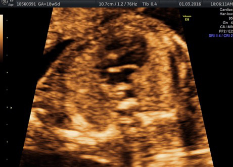
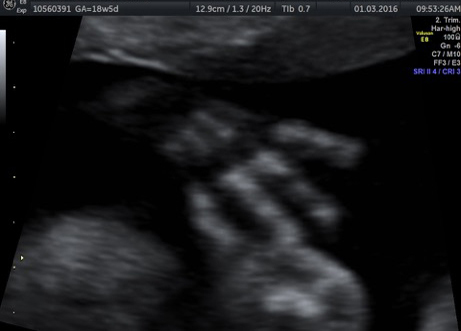
The doctor will point out your baby’s heartbeat and parts of his body, such as his face and hands, before looking at it in detail.
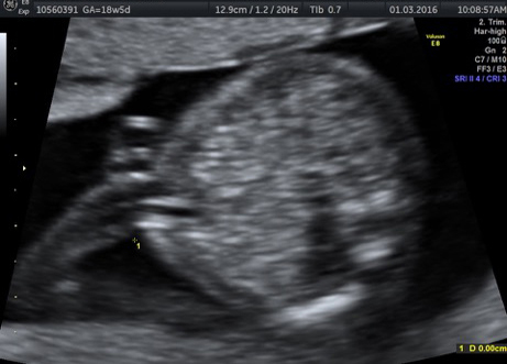
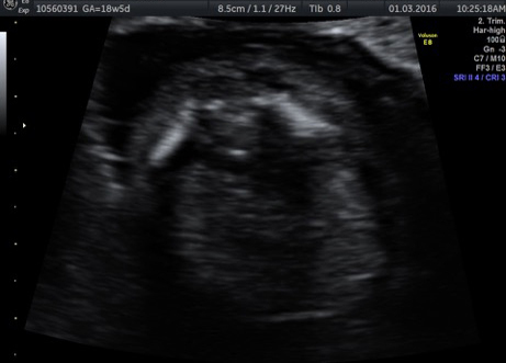
It may be hard for you to make out your baby’s organs, because the doctor will look at them in various angles and sections.
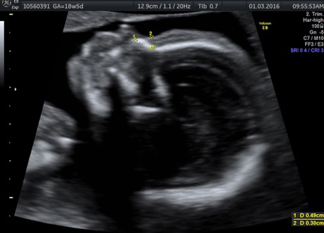
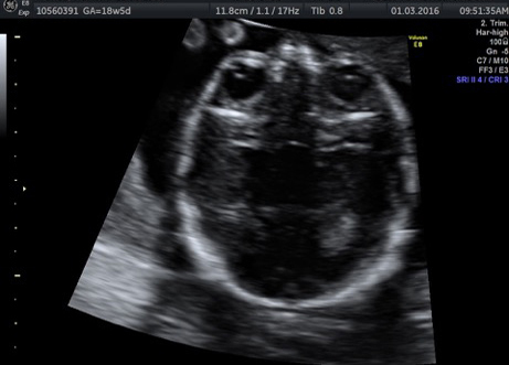
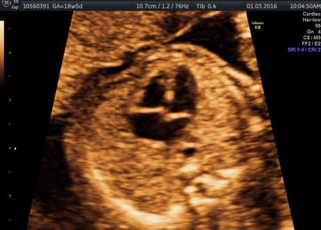
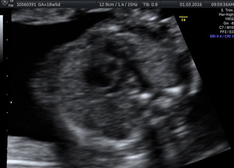
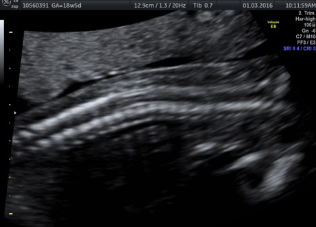
Your baby’s bones will appear white on the scan and its soft tissues look grey and speckled. The amniotic fluid surrounding your baby will look black.
Most serious abnormalities can be detected on a scan. However, it is not possible to see all problems and some will only be found after birth. Some conditions such as cerebral palsy and autism will never be seen on a scan.
The quality of the scan image depends on many factors, including the position of the baby and the size of the mother. For example, it will be more difficult to see the baby clearly if the mother is overweight. A poor image will affect our ability to see problems.
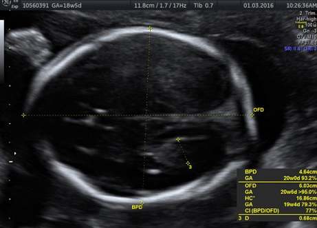
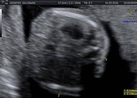
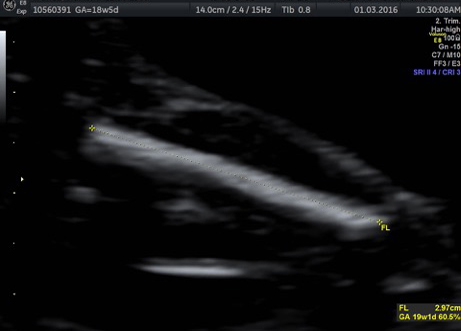
During the scan, the doctor will measure parts of your baby’s body, to see how well it is growing. The doctor will measure your baby’s:
- head circumference (HC)
- abdominal circumference (AC)
- femur or thigh bone (FL)
- humeris or arm bone
The measurements should match up to what’s expected for your baby, given its anticipated due date.
The doctor will look at the placenta (after birth). The placenta will be described as low if it reaches down to or covers the neck of your uterus (your cervix). If the placenta is low lying, you’ll have another scan in the eighth month to check its position. By then, it’s likely the placenta will have moved away from your cervix.
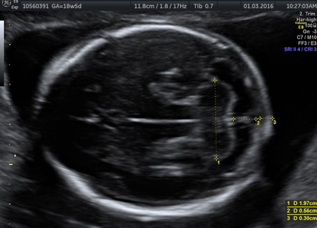
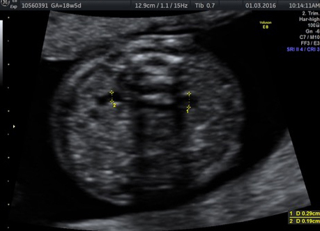
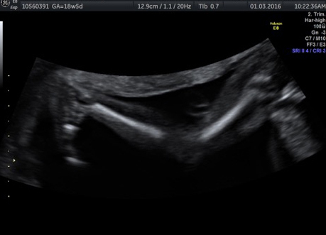
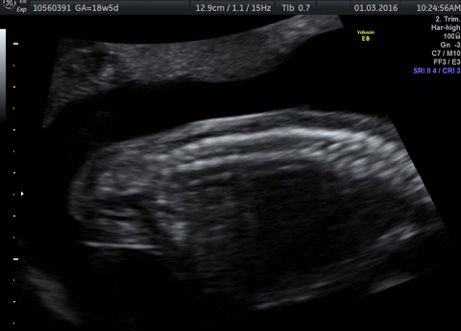
The doctor will then look at the baby’s head, face, spine, abdominal wall, heart, stomach, kidneys, umbilical cord, hands, feet, amniotic fluid in detail.
Below is a list of different types of congenital abnormality, and how likely scanning is to identify each problem.
| Problem | What the problem is | Chance of being seen |
|---|---|---|
| Spina bifida | Open spinal cord | >90% |
| Anencephaly | Absence of the top of the head | 99% |
| Hydrocephalus | *Excess fluid within the brain | 60% |
| Major congenital heart problems | 60% | |
| Diaphragmatic hernia | A defect in the muscle which separates the chest and abdomen | 60% |
| Exomphalos/gastroschisis | Defects of the abdominal wall | 90% |
| Major kidney problems | Missing or abnormal kidneys | 85% |
| Major limb abnormalities | Missing or abnormal limbs | 90% |
| Cerebral palsy | Spasticity | Never seen |
| Autism | Never seen | |
| Down syndrome | May be associated with heart and bowel problems | About 50-60% |
| * Many cases present late in pregnancy or even after birth | ||
This means that even if your scan is normal there is a small chance that your baby will still have a problem.
Some conditions, including certain heart defects and bowel obstructions, may not be seen until later in your pregnancy. Having your anomaly scan will most likely rule out all these conditions, as the vast majority of babies are born healthy.
This scan can also identify 50% to 60% of cases of Down syndrome, but the First Trimester Screening (FTS) test is definitely better for this. Because 30% to 50% of cases of Down syndrome appear normal on ultrasound, only an amniocentesis (testing the fluid surrounding the baby for its chromosomes) can give you this information for certain.
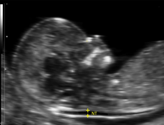
Sometimes babies with chromosomal abnormalities have signs called ultrasound markers. These include in order of importance thick skin behind the neck (nuchal fold), absent nasal bone, mild fluid within the ventricles of the brain, aberrant subclavian artery in the neck, occasionally short arms or legs, white spots in the baby’s heart or abdomen, or choroid plexus cysts in the brain.
While some babies with chromosomal abnormalities have these markers, it is important to remember that many normal babies also have these signs. The only way to diagnose or exclude a chromosomal problem for certain is to have an amniocentesis.
Most problems that need repeat scanning are not serious. About 15 per cent of scans will be done again for one reason or another.
The most common reason is that the doctor has not seen everything she needs to see. This may be because your baby is not lying in a good position, or you are overweight, in which case the scan should be repeated after a week or two.
If your doctor finds or suspects a problem, you will be told straight away.
If doctors suspect that your baby has a heart problem, you will be asked to come in a fetal echo. The fetal echo scan will take a detailed look at your baby’s heart.
If any scan reveals a serious problem, you will be given plenty of support to guide you through all the options. Although such serious problems are rare, some families are faced with the most difficult decision of all, whether to end the pregnancy.
Other problems may mean a baby needs surgery or treatment after birth, or even surgery while it is still in the uterus. There will be a whole range of people to support you through any difficult times, including obstetricians, pediatricians, pediatric surgeons, pediatric cardiologists/neurologists etc.
You could read the details of certain conditions that could be diagnosed on the home page of the website.