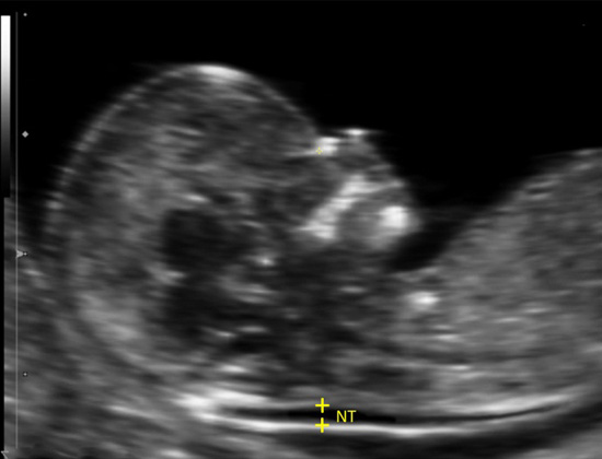


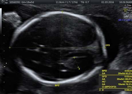
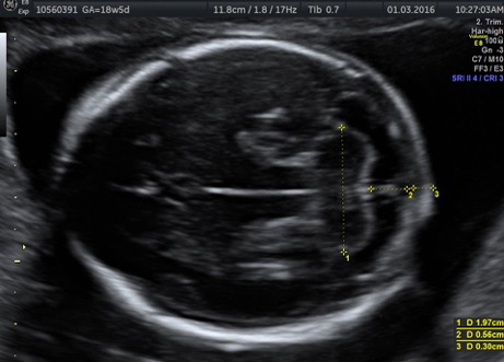
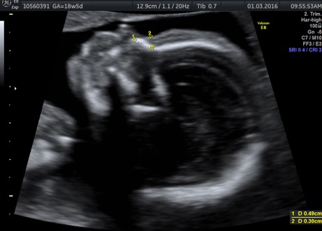
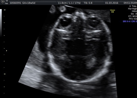

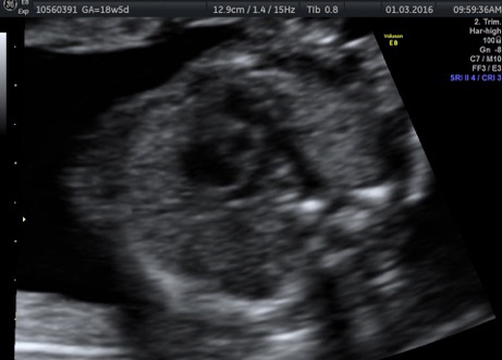
A Doppler ultrasound test uses reflected sound waves to see how blood flows through a blood vessel. It helps doctors evaluate blood flow through major arteries and veins, such as those of the arms, legs, and neck. It can show blocked or reduced flow of blood through narrow areas in the major arteries of the neck that could cause a stroke. It also can reveal blood clots in leg veins (deep vein thrombosis, or DVT) that could break loose and block blood flow to the lungs (pulmonary embolism). During pregnancy, Doppler ultrasound may be used to look at blood flow in an unborn baby (fetus) to check the health of the fetus.
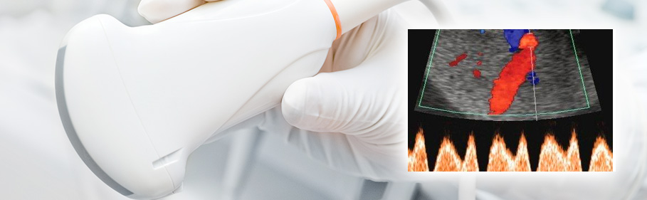
During Doppler ultrasound, a handheld instrument (transducer) is passed lightly over the skin above a blood vessel. The transducer sends and receives sound waves that are amplified through a microphone. The sound waves bounce off solid objects, including blood cells. The movement of blood cells causes a change in pitch of the reflected sound waves (called the Doppler effect). If there is no blood flow, the pitch does not change. Information from the reflected sound waves can be processed by a computer to provide graphs or pictures that represent the flow of blood through the blood vessels. These graphs or pictures can be saved for future review or evaluation.
“Bedside” or continuous wave Doppler.
This type uses the change in pitch of the sound waves to provide information about blood flow through a blood vessel. The doctor listens to the sounds produced by the transducer to evaluate the blood flow through an area that may be blocked or narrowed. This type of ultrasound can be done at the bedside in the hospital with a portable machine to provide a fast estimate of the extent of blood vessel damage or disease.
Duplex Doppler.
Duplex Doppler ultrasound uses standard ultrasound methods to produce a picture of a blood vessel and the surrounding organs. Also, a computer converts the Doppler sounds into a graph that gives information about the speed and direction of blood flow through the blood vessel being evaluated.
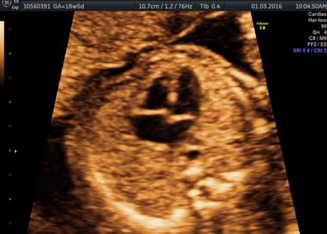
Color Doppler.
Color Doppler uses standard ultrasound methods to produce a picture of a blood vessel. Also, a computer converts the Doppler sounds into colors that are overlaid on the image of the blood vessel and that represent the speed and direction of blood flow through the vessel. Power Doppler is a special type of color Doppler. Power Doppler can get some images that are hard or impossible to get using standard color Doppler. Power Doppler is most commonly used to evaluate blood flow through vessels within solid organs.